Placental Cord Insertion Variations: From Normal Attachment to Abnormal Vessel Pathways
0% Complete
Course Overview
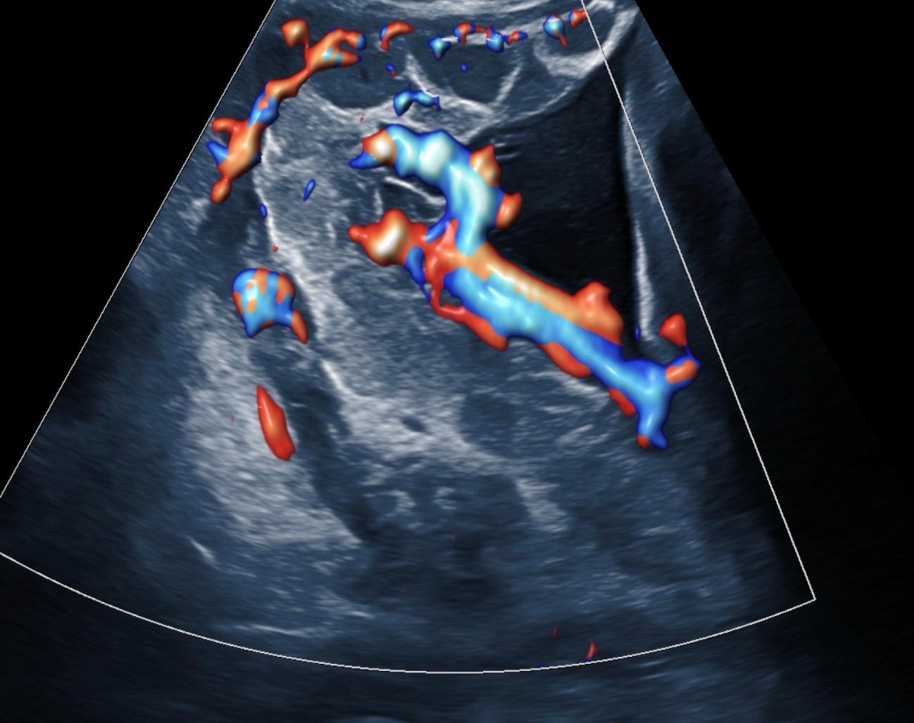
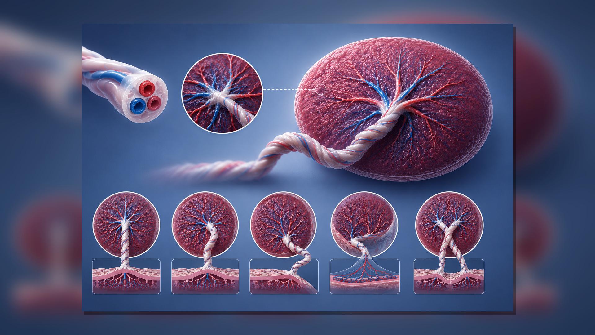
This course provides a focused review of placental cord insertion variations and their importance in obstetric ultrasound assessment. Learners will examine the sonographic features of central, eccentric/paracentral, marginal, velamentous, and furcate cord insertions, with emphasis on gray-scale recognition, color Doppler confirmation, and clinical significance.
The course highlights a systematic approach to evaluating the placental cord insertion site, including assessment of cord entry, placental edge relationship, exposed fetal vessels, and vessel mobility. Participants will review key imaging pitfalls and learn how to differentiate marginal cord insertion from velamentous cord insertion using ultrasound anatomy and Doppler findings.
Clinical relevance is emphasized throughout the course, particularly the association between abnormal cord insertion, fetal growth restriction, vasa previa risk, and the need for appropriate follow-up or referral. Learners will also review when transvaginal ultrasound with color Doppler is indicated to assess vessels near the internal cervical os.
By the end of this course, participants will be better prepared to recognize placental cord insertion variants, apply appropriate ultrasound techniques, and document findings using accurate, clinically meaningful terminology.
Course Content
Credits
* AMA PRA Category 1 Credit™ is designated for and awarded to physicians. Non-physician healthcare professionals, including physician assistants, nurse practitioners, and sonographers may participate in activities accredited for AMA PRA Category 1 Credit™; their respective credentialing and professional organizations independently recognize such participation toward their own continuing education requirements. Organizations that accept AMA PRA Category 1 Credit™-accredited courses toward non-physician CE include ARDMS, CCI, and Sonography Canada.
Course Details
- Duration
- 1.00 hour
- Released
- May 18, 2026
- Last Review
- May 18, 2026
- Expires
- May 18, 2029
Objectives
Define central, paracentral/eccentric, marginal, velamentous, and furcate placental cord insertion patterns.
Describe the sonographic findings that distinguish true parenchymal insertion from membrane-bound or unprotected vessels.
Apply a systematic gray-scale and color/power Doppler scanning protocol for PCI documentation at the anatomy scan.
Identify lower uterine segment findings that should trigger targeted transvaginal color Doppler evaluation for vasa previa.
Use structured reporting phrases that communicate location, distance, vessel protection, Doppler findings, and scan limitations.
Target Audience
Diagnostic medical sonography students
OB/GYN sonographers
Maternal-fetal medicine sonographers
Physicians, advanced practice providers, and interpreting clinicians involved in obstetric imaging
Faculty & Disclosure
Faculty
Lashell Jones RDMS, RDCS
Disclosure
Lashell Jones RDMS, RDCS discloses no such relationships exist
No one at IAME who had control over the planning or content of this activity has relationships with commercial interests.
Accreditation
The Institute for Advanced Medical Education is accredited by the Accreditation Council for Continuing Medical Education (ACCME) to provide continuing medical education for physicians.
The Institute for Advanced Medical Education designates this enduring material for a maximum of 1.00 AMA PRA Category 1 Credit™.
Physicians should only claim credit commensurate with the extent of their participation in the activity. Sonographers: These courses are accepted by the American Registry for Diagnostic Medical Sonography (ARDMS), Sonography Canada, Cardiovascular Credentialing International (CCI), and most other organizations.
Non physicians: AMA PRA Category 1 Credit™ is designated for and awarded to physicians. Non-physician healthcare professionals — including physician assistants, nurse practitioners, and others — may participate in activities accredited for AMA PRA Category 1 Credit™; their respective credentialing and professional organizations independently recognize such participation toward their own continuing education requirements.

Related Courses
Placenta Accreta Spectrum (PAS): Early Recognition, Reporting, and Referral in Obstetric Imaging