Exploring Hypoechoic Masses in Breast Imaging: A Comprehensive Review
Introduction
In the field of breast imaging, the identification and characterization of hypoechoic masses play a crucial role in the diagnosis and management of breast conditions. This comprehensive review aims to provide sonographers, doctors, nurse practitioners, physician assistants, and other healthcare professionals with a detailed understanding of hypoechoic masses in breast imaging.
Understanding Hypoechoic Masses
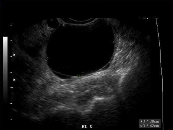
Hypoechoic masses refer to areas within the breast that appear darker or less bright on ultrasound imaging compared to the surrounding tissue. These masses can be benign or malignant and require careful evaluation to determine their nature accurately.
Diagnosis and Differential Diagnosis
Accurate diagnosis of hypoechoic masses requires a combination of imaging techniques, including ultrasound, mammography, and magnetic resonance imaging (MRI). Differential diagnosis involves considering various factors such as the shape, margins, presence of calcifications, and vascularity of the mass.
Role of CME in Breast Imaging
Continuing Medical Education (CME) is essential for healthcare professionals involved in breast imaging. CME activities provide opportunities to stay updated with the latest advancements and evidence-based practices in the field. Regular participation in CME courses and conferences ensures that healthcare professionals maintain their expertise and deliver the highest quality of care to patients.
Requirements for CME in Breast Imaging
The requirements for CME in breast imaging vary depending on the professional’s role and specialty. However, most healthcare professionals are required to complete a specific number of CME credits annually or within a designated timeframe. These credits can be earned through attending conferences, workshops, online courses, or participating in research and educational activities.
Importance of CME in Breast Imaging
CME is crucial in breast imaging as it allows healthcare professionals to update their knowledge and skills in the rapidly evolving field. It enables them to stay abreast of new technologies, imaging guidelines, and research findings, ensuring accurate and timely diagnosis of breast conditions. CME also promotes collaboration among healthcare professionals, fostering a multidisciplinary approach to patient care.
Conclusion
In conclusion, understanding hypoechoic masses in breast imaging is vital for healthcare professionals involved in the diagnosis and management of breast conditions. Continuous education through CME is essential to ensure healthcare professionals stay current with the latest advancements and provide optimal care to patients. By actively participating in CME activities, healthcare professionals can enhance their expertise and contribute to the advancement of breast imaging practices.


