Exploring the Link Between Hypoechoic Breast Masses and Breast Cancer
Introduction
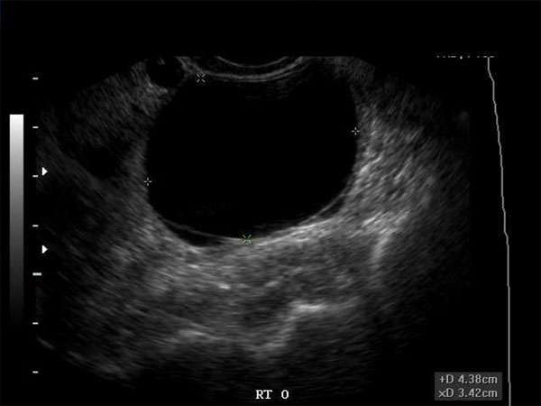
Hypoechoic breast masses are a common finding in ultrasound examinations. They refer to areas in the breast that appear darker or less reflective on the ultrasound image. While hypoechoic masses can have various causes, one important consideration is the potential link to breast cancer. This article aims to delve into the connection between hypoechoic breast masses and breast cancer, providing valuable insights for sonographers, doctors, and other healthcare professionals.
Hypoechoic Breast Masses and Breast Cancer
Research has shown that hypoechoic breast masses can be an indication of breast cancer. These masses often exhibit irregular borders, microlobulations, and spiculations on ultrasound images, which are characteristic features of malignant tumors. However, it is important to note that not all hypoechoic masses are cancerous, as benign conditions such as fibroadenomas can also present as hypoechoic masses.
Diagnostic Importance
Identifying hypoechoic breast masses and distinguishing between benign and malignant masses is crucial for early detection and appropriate management of breast cancer. Sonographers, radiologists, OB/GYNs, and other healthcare professionals involved in breast imaging play a critical role in this process. By accurately identifying and characterizing hypoechoic masses, they can aid in the early diagnosis of breast cancer, leading to improved patient outcomes.
Continuing Medical Education (CME) Requirements
Given the importance of accurate interpretation of breast ultrasound findings, healthcare professionals involved in breast imaging are encouraged to participate in Continuing Medical Education (CME) activities. CME programs provide opportunities for professionals to enhance their knowledge and skills in breast imaging, including the identification and characterization of hypoechoic masses. These educational activities help healthcare professionals stay updated with the latest advancements in breast imaging and improve their ability to detect and diagnose breast cancer.
Importance of CME
Continuing Medical Education is not only essential for maintaining professional competence but also for ensuring patient safety and quality healthcare. It allows healthcare professionals to stay abreast of emerging research, technological advancements, and best practices in their respective fields. CME activities specifically tailored to breast imaging equip professionals with the knowledge and expertise needed to make accurate assessments and provide appropriate patient care.
Conclusion
The link between hypoechoic breast masses and breast cancer highlights the significance of accurate identification and characterization of these masses in ultrasound imaging. Sonographers, doctors, and other healthcare professionals involved in breast imaging should prioritize their Continuing Medical Education to stay updated with the latest developments in breast imaging and improve patient outcomes. By continuously enhancing their skills and knowledge, healthcare professionals can ensure early detection and appropriate management of breast cancer.


