Management Strategies for Hypoechoic Breast Masses: A Practical Approach
Introduction
Hypoechoic breast masses are a common finding in clinical practice, often requiring further evaluation and management. In this article, we will discuss practical management strategies for hypoechoic breast masses, providing valuable insights for sonographers, doctors, nurse practitioners, physician assistants, and other healthcare professionals.
Understanding Hypoechoic Breast Masses
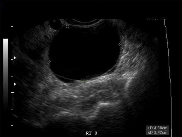
Hypoechoic breast masses refer to lesions that appear darker on ultrasound images compared to the surrounding breast tissue. These masses can be benign or malignant and require careful assessment to determine the appropriate management approach. Sonographers and healthcare professionals involved in breast imaging should be familiar with the characteristics of hypoechoic breast masses.
Diagnostic Evaluation
When hypoechoic breast masses are detected, a comprehensive diagnostic evaluation is crucial. This may include additional imaging modalities such as mammography, magnetic resonance imaging (MRI), or biopsy procedures. Doctors, radiologists, and other specialists play a vital role in interpreting these results and determining the next steps in management.
Management Strategies
The management of hypoechoic breast masses depends on several factors, including the patient’s age, medical history, imaging findings, and biopsy results. Here are some practical strategies for managing hypoechoic breast masses:
1. Close Surveillance
In cases where the mass is deemed low-risk or highly likely to be benign, close surveillance with regular follow-up imaging may be recommended. This approach ensures that any changes in the mass can be closely monitored and acted upon if necessary.
2. Biopsy and Histopathological Evaluation
If the hypoechoic breast mass is suspicious or shows concerning features on imaging, a biopsy may be necessary to obtain tissue samples for histopathological evaluation. This helps in accurately diagnosing the mass and determining the appropriate treatment plan.
3. Multidisciplinary Approach
Collaboration among healthcare professionals is essential in managing hypoechoic breast masses. Radiologists, OB/GYNs, vascular surgeons, and other specialists should work together to discuss and decide on the most appropriate management strategy for each individual case.
The Importance of Continuing Medical Education (CME)
Continuing Medical Education (CME) plays a vital role in ensuring that healthcare professionals stay updated with the latest advancements and best practices in their respective fields. Sonographers, doctors, nurse practitioners, physician assistants, and other healthcare professionals involved in breast imaging should actively participate in CME activities related to breast pathology and management.
Requirements for CME
Each medical specialty has specific requirements for CME credits that healthcare professionals must fulfill to maintain their licensure and stay up-to-date with current knowledge. It is crucial for sonographers, doctors, and other healthcare professionals to be aware of their specialty’s CME requirements and actively engage in relevant educational activities.
Conclusion
Management strategies for hypoechoic breast masses require a practical, multidisciplinary approach involving various healthcare professionals. Sonographers, doctors, nurse practitioners, and physician assistants should stay updated through continuing medical education to ensure they provide the best possible care to their patients. By following evidence-based approaches and participating in CME activities, healthcare professionals can effectively manage hypoechoic breast masses and improve patient outcomes.

