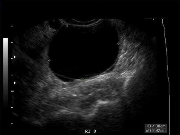
Ultrasound Imaging of Hypoechoic Breast Masses: An Essential Tool for Early Detection
Introduction
Ultrasound imaging plays a crucial role in the early detection and diagnosis of breast abnormalities, particularly hypoechoic breast masses. This article aims to highlight the significance of ultrasound imaging in identifying and evaluating these masses, and its importance in improving patient outcomes.
The Role of Ultrasound Imaging
Ultrasound imaging is a non-invasive and radiation-free modality that utilizes high-frequency sound waves to produce detailed images of internal structures. In the case of hypoechoic breast masses, ultrasound can provide valuable information regarding their characteristics, size, location, and vascularity.
Early Detection and Diagnosis
Early detection of breast abnormalities, including hypoechoic masses, is crucial for timely intervention and improved prognosis. Ultrasound imaging allows for the detection of masses that may not be visible on mammography or other imaging modalities, especially in women with dense breast tissue.
Characterization of Hypoechoic Masses
Ultrasound imaging helps in characterizing hypoechoic breast masses by assessing their shape, margins, echogenicity, and internal features. These characteristics aid in differentiating benign masses from potentially malignant ones, guiding further diagnostic interventions.
Importance of Continuing Medical Education (CME)
For sonographers, doctors, nurse practitioners, physician assistants, and other healthcare professionals involved in breast imaging, staying updated with the latest advancements and guidelines is essential. Continuing Medical Education (CME) provides an avenue for professionals to enhance their skills, knowledge, and expertise in ultrasound imaging of breast masses.
Requirements for CME
Various professional organizations, such as the American College of Radiology (ACR) and the American Institute of Ultrasound in Medicine (AIUM), offer CME programs focused on breast imaging. These programs often include courses, conferences, workshops, and online resources that cover the latest techniques, guidelines, and research in the field.
Benefits of CME
Engaging in CME activities allows healthcare professionals to stay current with the evolving field of breast imaging. It fosters continuous improvement in clinical practice, enhances diagnostic accuracy, and promotes patient safety. Moreover, CME activities provide opportunities for networking, collaboration, and sharing of best practices among professionals.
Conclusion
Ultrasound imaging is an essential tool for the early detection and diagnosis of hypoechoic breast masses. Its ability to accurately characterize these masses aids in determining appropriate management strategies. Additionally, engaging in CME activities ensures that healthcare professionals are equipped with the necessary knowledge and skills to provide the highest quality of care in breast imaging.


