Understanding Hypoechoic Breast Masses: Causes, Diagnosis, and Treatment
Introduction
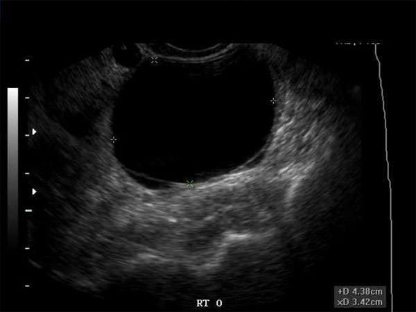
Hypoechoic breast masses are a common finding in medical imaging, particularly in ultrasound examinations. They refer to areas in the breast that appear darker or less echogenic than the surrounding tissue. Understanding the causes, diagnosis, and treatment of hypoechoic breast masses is crucial for healthcare professionals, including sonographers, doctors, nurse practitioners, and physician assistants.
Causes of Hypoechoic Breast Masses
There are various causes of hypoechoic breast masses, including:
- Fibroadenomas: These are benign solid masses commonly found in young women. They are usually well-defined and have a hypoechoic appearance.
- Breast cysts: Fluid-filled sacs within the breast tissue can appear hypoechoic on ultrasound. They are often round or oval in shape.
- Breast cancer: Some breast cancers manifest as hypoechoic masses. However, it is important to note that not all hypoechoic masses indicate cancer.
- Inflammation and infection: Conditions like mastitis or abscesses can cause hypoechoic areas in the breast.
Diagnosis of Hypoechoic Breast Masses
Accurate diagnosis of hypoechoic breast masses requires imaging techniques and sometimes additional procedures. The following steps are commonly taken:
- Ultrasound imaging: Ultrasound is the primary tool for visualizing and characterizing hypoechoic breast masses. It helps determine the size, shape, and composition of the mass.
- Mammography: Mammograms may be performed to provide additional information about the mass, especially in women over 40 years old.
- Magnetic Resonance Imaging (MRI): In certain cases, an MRI may be ordered to obtain more detailed images of the breast and surrounding tissues.
- Biopsy: A biopsy may be necessary to confirm the nature of the mass. This involves extracting a sample of tissue for examination under a microscope.
Treatment Options
The appropriate treatment for hypoechoic breast masses depends on their underlying cause. The options include:
- Observation: Many benign hypoechoic masses, such as fibroadenomas, can be safely observed without immediate intervention.
- Aspiration or drainage: Fluid-filled cysts can often be drained using a needle and syringe. This procedure provides immediate relief and helps confirm the cystic nature of the mass.
- Surgical excision: In cases where malignancy is suspected or confirmed, surgical removal of the mass may be necessary.
- Medical management: Breast infections and inflammations are typically treated with antibiotics or anti-inflammatory medications.
Importance of Continuing Medical Education (CME)
For healthcare professionals involved in breast imaging and the diagnosis of hypoechoic breast masses, Continuing Medical Education (CME) is crucial. CME courses and activities provide opportunities for professionals to stay updated with the latest advancements, guidelines, and techniques in breast imaging. It helps them enhance their diagnostic skills and improve patient outcomes.
CME requirements vary by profession and specialty, but they often involve completing a certain number of accredited educational activities and obtaining a specific number of credits or hours. These requirements ensure that healthcare professionals maintain their knowledge and skills throughout their careers.
By participating in CME, sonographers, doctors, nurse practitioners, physician assistants, and other healthcare professionals can stay up-to-date with the rapidly evolving field of breast imaging. It allows them to provide the best possible care for patients with hypoechoic breast masses and make well-informed diagnostic and treatment decisions.
In conclusion, understanding hypoechoic breast masses is essential for healthcare professionals involved in breast imaging. By staying updated through continuing medical education, these professionals can effectively diagnose and manage hypoechoic breast masses, ultimately improving patient outcomes.


