Understanding Hypoechoic Breast Masses: Causes, Symptoms, and Treatment
Introduction:
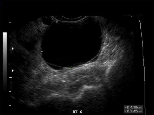
Hypoechoic breast masses are a common finding in clinical practice, often detected during breast ultrasound examinations. These masses appear darker than the surrounding breast tissue on ultrasound images, raising concerns about potential malignancy. It is crucial for healthcare professionals, including sonographers, doctors, nurse practitioners, and physician assistants, to understand the causes, symptoms, and appropriate treatment options for hypoechoic breast masses. Continuous medical education (CME) plays a vital role in ensuring healthcare professionals stay updated with the latest advancements in this field.
1. Causes of Hypoechoic Breast Masses:
Hypoechoic breast masses can have various causes, including:
– Benign Breast Conditions: Fibroadenomas, cysts, and intramammary lymph nodes are common benign causes of hypoechoic breast masses.
– Breast Cancer: Hypoechoic masses can be indicative of breast cancer, particularly invasive ductal carcinoma. However, not all hypoechoic masses are cancerous, highlighting the importance of further evaluation.
– Infection or Inflammation: Mastitis or abscesses can also present as hypoechoic masses, necessitating appropriate diagnosis and treatment.
– Trauma: In some cases, hypoechoic masses may be the result of previous trauma, such as a hematoma or fat necrosis.
2. Symptoms and Clinical Presentation:
Hypoechoic breast masses may or may not be associated with symptoms. Some common signs and symptoms include:
– Palpable lump: Patients may notice a lump or mass during self-examination or clinical breast examination.
– Pain or tenderness: Hypoechoic masses, especially those associated with infection or inflammation, can cause localized pain or tenderness.
– Skin changes: In some cases, hypoechoic masses may be accompanied by skin changes, such as redness, warmth, or dimpling.
– Nipple discharge: Although not specific to hypoechoic masses, the presence of nipple discharge should be evaluated further.
3. Treatment Options:
The treatment approach for hypoechoic breast masses depends on the underlying cause. Some common treatment options include:
– Observation: If the mass is determined to be benign and does not pose immediate concerns, periodic monitoring may be recommended.
– Biopsy: In cases where malignancy is suspected or cannot be ruled out, a biopsy may be necessary to obtain tissue for further evaluation.
– Medications: Antibiotics or anti-inflammatory medications may be prescribed for hypoechoic masses associated with infection or inflammation.
– Surgical Intervention: Malignant masses or certain benign masses may require surgical excision for definitive treatment.
4. Importance of Continuous Medical Education (CME):
For healthcare professionals, including sonographers, doctors, nurse practitioners, and physician assistants, staying updated with the latest knowledge and techniques is crucial. Continuous medical education (CME) offers opportunities to enhance professional competence, improve patient care, and meet licensing requirements. It ensures healthcare professionals are equipped with the necessary skills and knowledge to accurately diagnose and manage hypoechoic breast masses.
Conclusion:
Understanding the causes, symptoms, and treatment options for hypoechoic breast masses is essential for healthcare professionals involved in breast imaging and patient care. Continuous medical education (CME) plays a vital role in ensuring healthcare professionals stay up-to-date with the latest advancements and standards in managing these masses. By staying informed and maintaining their expertise, healthcare professionals can provide optimal care to patients with hypoechoic breast masses.


