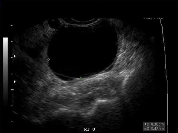
Differentiating Hypoechoic Breast Masses: Benign vs. Malignant Lesions
Introduction
When it comes to breast health, early detection and accurate diagnosis of breast masses are crucial in determining the appropriate course of treatment. Sonographers, doctors, nurse practitioners, and other healthcare professionals play a vital role in identifying and differentiating between benign and malignant lesions in the breast. This article aims to provide an overview of the differentiation process and emphasize the significance of continuing medical education (CME) in this field.
Understanding Hypoechoic Breast Masses
Hypoechoic breast masses refer to areas within the breast that appear darker or less reflective on ultrasound imaging compared to the surrounding tissue. These masses can be either benign or malignant, making it essential to accurately differentiate between the two to guide patient management decisions.
Differentiating Benign Lesions
Benign breast masses are non-cancerous growths that do not pose a significant health risk. Common benign lesions include cysts, fibroadenomas, and intramammary lymph nodes. Sonographers and healthcare professionals should consider certain characteristics when assessing these lesions:
- Smooth borders
- Uniform internal echoes
- Well-defined oval or round shapes
- Presence of posterior enhancement (increased echoes behind the mass)
Differentiating Malignant Lesions
Malignant breast lesions indicate the presence of cancer cells and require immediate attention. Common malignant lesions include invasive ductal carcinoma, invasive lobular carcinoma, and ductal carcinoma in situ. Key features of malignant lesions include:
- Irregular borders
- Microlobulations or spiculations
- Heterogeneous internal echoes
- Increased vascularity (seen through Doppler imaging)
The Importance of Continuing Medical Education (CME)
In the rapidly evolving field of breast imaging and diagnosis, staying updated with the latest advancements is crucial. CME programs provide healthcare professionals with the opportunity to enhance their knowledge and skills, ensuring accurate and timely diagnosis of breast masses. Sonographers, doctors, nurse practitioners, and other healthcare professionals should actively participate in CME activities to:
- Stay informed about emerging technologies and techniques
- Improve accuracy in differentiating between benign and malignant lesions
- Understand the latest guidelines and recommendations for breast imaging
- Enhance patient care and outcomes
Conclusion
Accurate differentiation between benign and malignant breast masses is essential for appropriate patient management. Sonographers, doctors, nurse practitioners, and other healthcare professionals should continuously update their knowledge and skills through CME programs to provide the best possible care to their patients. By staying informed and proficient in the latest advancements, healthcare professionals can make a significant impact on breast health outcomes.


