Hypoechoic Lesions in Breast Ultrasound: Evaluation and Diagnostic Challenges
Introduction
Hypoechoic lesions in breast ultrasound are a common finding during diagnostic evaluations. These lesions often pose diagnostic challenges for sonographers, doctors, and other healthcare professionals. Understanding the evaluation process and the importance of continuing medical education (CME) is crucial in accurately diagnosing and managing these lesions.
Understanding Hypoechoic Lesions
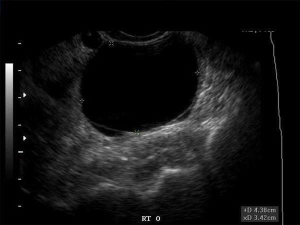
Hypoechoic lesions appear as dark or hypoechoic areas on ultrasound images, indicating that they reflect less sound waves compared to the surrounding tissue. In breast ultrasound, these lesions can represent various abnormalities, including cysts, fibroadenomas, or even malignant tumors. Accurate evaluation and diagnosis are essential to determine the appropriate course of action.
Diagnostic Challenges
Evaluating hypoechoic lesions can be challenging due to their varied appearances and potential overlap with normal breast tissue. Distinguishing between benign and malignant lesions can be particularly difficult, requiring comprehensive knowledge and expertise in breast ultrasound interpretation. Misinterpretation can lead to unnecessary biopsies or missed diagnoses, emphasizing the need for continuing medical education.
Importance of Continuing Medical Education (CME)
Continuing medical education plays a vital role in ensuring healthcare professionals stay up-to-date with the latest advancements and best practices in their respective fields. For sonographers, radiologists, OB/GYNs, vascular surgeons, and other healthcare professionals involved in breast ultrasound, CME courses provide opportunities to enhance their knowledge and skills in evaluating hypoechoic lesions.
Requirements for CME
Healthcare professionals should actively pursue CME activities to meet the requirements set by their respective medical boards and professional organizations. In the field of breast ultrasound, CME courses typically cover topics such as breast anatomy, lesion characterization, imaging techniques, and biopsy guidance. Regular participation in CME ensures that healthcare professionals maintain their competence and deliver optimal patient care.
Conclusion
Evaluating and diagnosing hypoechoic lesions in breast ultrasound require a comprehensive understanding of the nuances and challenges associated with these lesions. Sonographers, doctors, and other healthcare professionals must prioritize continuing medical education to stay updated with the latest developments in breast ultrasound and maintain their competency. By doing so, they can provide accurate diagnoses and improve patient outcomes.


