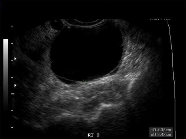
The Role of Ultrasound in Detecting Hypoechoic Breast Masses
Introduction
Ultrasound has become an invaluable tool in the early detection and diagnosis of various medical conditions. In the field of breast imaging, ultrasound plays a crucial role in identifying hypoechoic breast masses. This article aims to highlight the significance of ultrasound in detecting hypoechoic breast masses and its importance in the healthcare industry.
What are Hypoechoic Breast Masses?
Hypoechoic breast masses refer to areas within the breast tissue that appear darker or hypoechoic compared to the surrounding tissue on an ultrasound scan. These masses can be either benign or malignant and require further evaluation to determine their nature.
The Role of Ultrasound in Detection
Ultrasound imaging is widely used as a non-invasive method to examine breast tissue and detect abnormalities. When it comes to hypoechoic breast masses, ultrasound can provide detailed images that help differentiate between benign and malignant masses. This imaging technique allows healthcare professionals to identify the size, shape, and location of the mass, aiding in the diagnosis and treatment planning process.
Benefits of Ultrasound in Hypoechoic Breast Mass Detection
Ultrasound offers several advantages in the detection of hypoechoic breast masses:
-
- Non-ionizing radiation: Unlike mammography, ultrasound does not use ionizing radiation, making it a safe imaging option, especially for younger patients.
-
- High sensitivity: Ultrasound has proven to be highly sensitive in detecting breast abnormalities, including hypoechoic masses.
-
- Real-time imaging: With ultrasound, healthcare professionals can visualize the breast tissue in real-time, allowing for immediate evaluation and intervention if necessary.
Continuing Medical Education (CME) Requirements
For healthcare professionals involved in breast imaging, maintaining their knowledge and skills through continuing medical education (CME) is vital. CME ensures that practitioners stay up to date with the latest advancements and techniques in breast imaging, including the use of ultrasound for detecting hypoechoic breast masses.
The Importance of CME
CME plays a crucial role in enhancing patient care and improving diagnostic accuracy. By attending CME activities, healthcare professionals can learn about new technologies, research findings, and best practices in breast imaging. This knowledge empowers them to provide more accurate diagnoses, leading to better patient outcomes.
Conclusion
Ultrasound is a valuable tool in the detection and characterization of hypoechoic breast masses. Its non-invasive nature, high sensitivity, and real-time imaging capabilities make it an essential modality for healthcare professionals involved in breast imaging. Continuing medical education ensures that practitioners stay updated with the latest techniques and advancements in ultrasound imaging, further enhancing the accuracy and quality of patient care in the detection of hypoechoic breast masses.


