Exploring Hypoechoic Breast Lesions: A Comprehensive Guide for Women
Introduction
Hypoechoic breast lesions are a common finding during breast ultrasound examinations. While they can be concerning, it’s important to understand that not all hypoechoic lesions are cancerous. In this comprehensive guide, we will explore what hypoechoic breast lesions are, their causes, and the importance of early detection and diagnosis.
Understanding Hypoechoic Breast Lesions
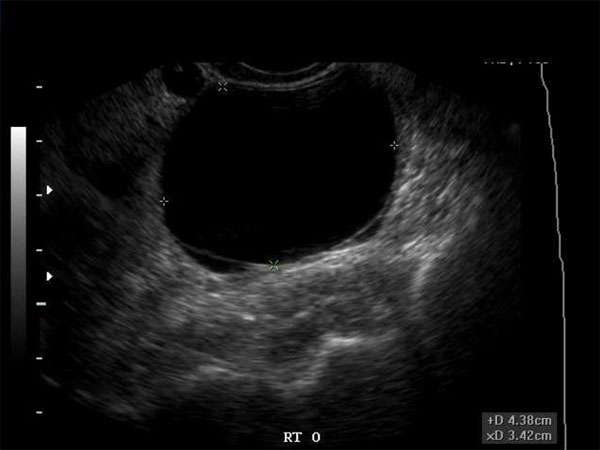
Hypoechoic breast lesions refer to areas in the breast that appear darker or black on ultrasound images. This darkness indicates that the tissue is reflecting fewer sound waves compared to the surrounding breast tissue. While hypoechoic lesions can occur in both benign and malignant conditions, further evaluation is necessary to determine their nature.
Common Causes of Hypoechoic Breast Lesions
There are several potential causes of hypoechoic breast lesions, including:
- Fibroadenomas: These are benign tumors that often present as hypoechoic lesions on ultrasound.
- Cysts: Fluid-filled sacs can also appear as hypoechoic lesions.
- Granulomas: Inflammatory conditions like tuberculosis or sarcoidosis can cause hypoechoic lesions.
- Breast cancer: Malignant tumors can present as hypoechoic lesions, but further evaluation is necessary for accurate diagnosis.
The Importance of Early Detection
Early detection of breast lesions, whether benign or malignant, is crucial for timely and appropriate management. Regular breast self-exams and routine mammograms are essential for identifying any abnormalities. Hypoechoic lesions detected during ultrasound examinations should be further evaluated to determine their nature and potential risks.
The Role of Continuing Medical Education (CME)
For sonographers, doctors, nurse practitioners, physician assistants, and other healthcare professionals, staying updated with the latest advancements in breast imaging is vital. Continuing Medical Education (CME) courses provide opportunities to enhance knowledge and skills in breast ultrasound and other diagnostic modalities. These courses cover topics such as identifying and characterizing hypoechoic breast lesions, interpreting ultrasound images, and understanding the latest guidelines for breast cancer screening.
Requirements for CME
The requirements for CME vary depending on the profession and specialization. However, most healthcare professionals are required to complete a certain number of CME credits annually or within a specified time frame. These credits can be earned through attending conferences, workshops, online courses, or participating in research and educational activities. It is important for healthcare professionals to fulfill their CME obligations to ensure they are providing the highest standard of care to their patients.
Conclusion
In conclusion, hypoechoic breast lesions are a common finding during breast ultrasound examinations. While they can be concerning, not all hypoechoic lesions are cancerous. Early detection and accurate diagnosis are crucial for appropriate management. Healthcare professionals should prioritize continuing medical education to stay updated with the latest advancements in breast imaging and ensure the best possible care for their patients.
Keywords: hypoechoic breast lesions, breast ultrasound, early detection, continuing medical education, CME requirements


