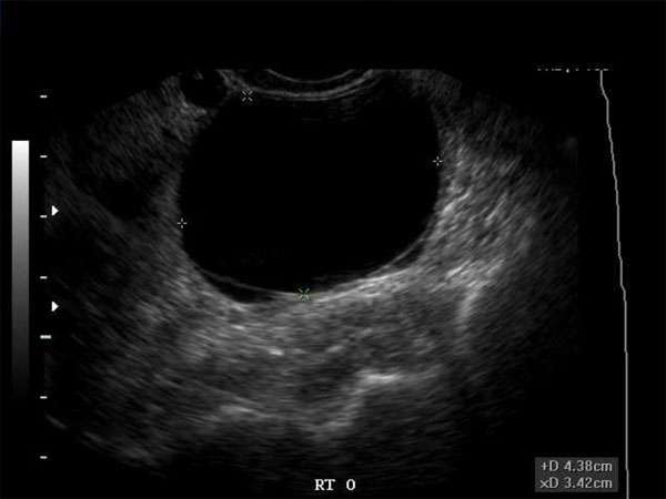
Hypoechoic Breast Masses: What They Mean and How to Identify Them
Introduction
Hypoechoic breast masses are a common finding during breast ultrasound examinations. It is important for sonographers, doctors, and other healthcare professionals to understand what these masses mean and how to accurately identify them.
Understanding Hypoechoic Breast Masses
Hypoechoic breast masses appear darker on ultrasound images compared to the surrounding breast tissue. They are often associated with solid lesions and can indicate the presence of a tumor or other abnormalities.
Identifying Hypoechoic Breast Masses
To identify hypoechoic breast masses, sonographers and healthcare professionals should follow a systematic approach:
1. Patient History and Clinical Examination
Obtain a detailed patient history and perform a thorough physical examination. This information can provide important clues about the nature of the breast mass.
2. Breast Ultrasound
Perform a breast ultrasound to visualize the breast tissue and identify any suspicious masses. Hypoechoic lesions should be carefully evaluated for shape, margins, and other characteristics.
3. Use of Doppler Imaging
Doppler imaging can help assess the vascularity of the hypoechoic mass. A highly vascularized mass may indicate a more aggressive tumor.
4. Comparison with Previous Imaging
If the patient has had previous mammograms or ultrasounds, compare the current images with the previous ones to track any changes in the hypoechoic mass over time.
Importance of Continuing Medical Education (CME)
For healthcare professionals, including sonographers, radiologists, OB/GYNs, and other specialists, staying up-to-date with the latest advancements and knowledge is crucial. Continuing Medical Education (CME) ensures that healthcare professionals maintain and enhance their competency in their respective fields.
Requirements for CME
The specific requirements for CME credits vary depending on the profession and country. However, most healthcare professionals need to complete a certain number of CME hours or credits within a specified time period. This can involve attending conferences, workshops, online courses, or participating in research and educational activities.
Benefits of CME
CME offers several benefits for healthcare professionals:
- Keeps professionals updated with the latest research and advancements in their field.
- Improves diagnostic and treatment skills, leading to better patient outcomes.
- Enhances communication and collaboration among healthcare teams.
- Provides opportunities for networking and knowledge sharing.
- Helps fulfill licensing and certification requirements.
Conclusion
Hypoechoic breast masses can indicate the presence of tumors or abnormalities. Sonographers, doctors, and other healthcare professionals should be well-versed in identifying and evaluating these masses during breast ultrasound examinations. Additionally, staying updated through Continuing Medical Education (CME) is essential to maintain competence and deliver the highest quality of care.


